
From the region
If you are within 100 kilometres, please book your appointment directly — by phone or via the online booking calendar. Bring your medical records with you; we will discuss everything else on site.
Conservative therapy recognises three levels at which it intervenes. The medication acts chemically. Physiotherapy, manual therapy and injections act mechanically. Surgery changes the structure. If nothing is achieved with this, the path usually ends.
Tissue has a fourth level: an electricalone. Cells align themselves with voltage gradients, wounds generate measurable currents, and cells change their migration direction in a field to close the wound. This is exactly where Axomera comes in — with the goal of influencing inflammation and regeneration where the three known levels have been exhausted.
„Modulating electrical cell signals may lead to a paradigm shift in the treatment of pain and inflammation. To my knowledge the Axomera therapy is the first to translate this into clinical reality.“
„Modulating electrical cell signals can lead to a paradigm shift in the treatment of pain and inflammation. To my knowledge, Axomera therapy is the first to translate this into clinical practice.“
Scientific co-author of the Axomera foundation publication (Molsberger & McCaig, 2018); co-author Nature 2006;442(7101):457–460.
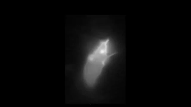
The animation is loaded from axomera.com only after you consent to external content.
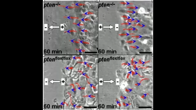
Every cell carries a voltage across its membrane. Injured or diseased tissue creates measurable electrical voltage gradients, along which cells align and migrate — cell biology calls this galvanotaxis. This basic principle has been described and molecularly explained since the 1970s: in 2006, a study in Natureshowed that electrical signals control wound healing via the same signalling pathway as the body's own messengers (PI3Kγ/PTEN).
Evidence: McCaig et al., Physiological Reviews 2005; Zhao et al., Nature 2006; Song et al., PNAS 2002. The complete citations are listed below in the literature list.
These foundations prove the principle — that electric fields influence cell migration, inflammation and regeneration. They provide the rationale for the working hypothesis of Axomera therapy, not proof of efficacy.
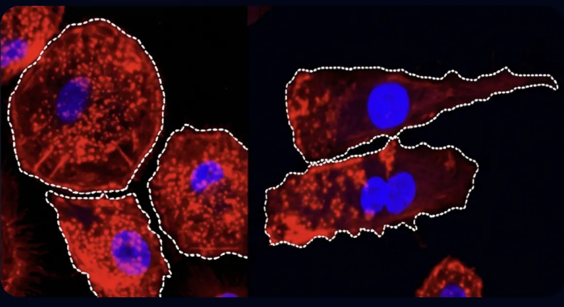
The underlying works (Zhao et al., Nature 2006; Song et al., PNAS 2002) show time-lapse microscopy in which cells align their migration direction according to an applied electric field.
Painfully irritated tissue is locally acidic. In active myofascial pain points, measurably more protons and inflammatory mediators can be found — substance P, CGRP, bradykinin, TNF-α, interleukin-1β — and a lower pH value than in healthy tissue; demonstrated via microdialysis directly in the muscle (Shah et al., 2005/2008). This acidification activates pain fibres and changes the electrical properties of the tissue.
Inflammatory cells, in turn, react to weak electric fields: even fields in the magnitude of the body's own currents steer the migration of macrophages and change their secretion of messenger substances (Hoare et al., 2016).
Axomera targets precisely this electrical level — one level deeper than medication, manual therapy and injections. Via the cathodic probe, the local acidosis is buffered and a field is generated that attracts repair cells; the goal is to modulate the inflammatory response and trigger regeneration (Molsberger & McCaig, 2018).
The animation is loaded from axomera.com only after you consent to external content.
More precisely: it is not the sensation that creates inflammation, but the activity of the pain fibre itself.
Inflammatory mediators and protons lower the excitation threshold of nociceptors — this is the evidence already mentioned in section 02 (microdialysis at active myofascial pain points). The mechanism behind this runs, among other things, via acid-sensing ion channels (ASIC) at the nerve ending: with a locally lowered pH value, these channels open and lower the irritation threshold of the fibre (Ugawa et al., 2005 — number 06 below). Thus, chemically inflamed tissue becomes electrically more excitable.
However, the path also works the other way around. Nociceptive C-fibres have, in addition to their afferent (signalling) function, also an efferent function. If an action potential travels outwards, it can, via the axon reflex — the back-propagation of electrical excitation into neighbouring branches of the same fibre without passing through the spinal cord — also release CGRP and substance P where no external stimulus was present. The consequence: vasodilation, plasma extravasation (leakage of blood plasma from vessels into tissue) and mast cell activation (Sann & Pierau, 1998 — number 31). This is exactly where the circle closes — and this is the reason why an intervention at a single point theoretically acts in both directions. Clinically, this feedback loop is one of the mechanisms through which acute pain can turn into chronic pain.
The interaction is not a law of nature that applies equally to every tissue. Depending on the structure, inflammation, disturbed remodelling or neither of the two predominates.
Neurogenic inflammation is the standard term for this mechanism — well-proven in animal experiments (Chiu, von Hehn & Woolf, 2012 — number 30 below). The direct translation of this model to human migraine is still being discussed in the specialist literature.
Low-grade synovitis (inflammation of the joint lining) is regularly detectable. The cartilage itself is nerve-free and cannot report pain — the pain comes from the synovia, bone marrow and joint capsule.
The compression of a healthy nerve root creates tingling and numbness, but no chronic pain; persistent ectopic (arising in the wrong place) electrical discharge only arises after previous damage to the root. Additionally, the leaked disc material itself has a chemical inflammatory effect. The circle applies here — but the mechanics alone are not enough as an explanation.
In load-bearing sections of chronically altered tendons, acute inflammatory cells are largely absent; a disrupted collagen turnover without a classic inflammatory response predominates (Khan et al., 2002 — number 28 below). This position is not undisputed: Rees, Stride & Scott (2014 — number 29) argue that inflammation does play a role in early and sub-sections of chronic tendinopathies. Both sides are presented here; neither is resolved.
An example of how clinical terminology can be misleading: "Bursitis trochanterica" suggests bursitis, whereas gluteal tendon tendinopathy is usually the actual cause; independent bursal inflammation is a minority finding (Klontzas & Karantanas, 2014 — number 32 below).
Nociplastic pain According to the IASP definition (introduced 2017, clinical criteria and grading 2021: Kosek et al. — number 33 below), this denotes pain arising from altered pain processing without clear evidence of tissue damage or inflammation. The approach described here does not explicitly apply to these cases.
If both directions are effective, an intervention at the electrical level is not determined by one direction alone. However, where tissue remodeling rather than inflammation is at the forefront — such as in chronic tendon issues — the rationale for using Axomera is different. This is precisely why the choice of words on the indication pages varies depending on the tissue. This section explains the underlying rationale — it makes no statement regarding efficacy.
Some animals regenerate entire body parts. The axolotl replaces limbs, and the frog begins leg regeneration following minute currents (Borgens et al., 1977). In the planarian worm, bioelectric voltage patterns even govern where the head and tail form — research by Michael Levin's group has shown that altered voltage can rewrite the blueprint (Durant et al., 2017). This research demonstrates: bioelectric control is a fundamental principle of living organisms.
Evidence: Borgens, Vanable & Jaffe, J. Exp. Zool. 1977; Durant et al., Biophysical Journal 2017; popular science summary in Spektrum der Wissenschaft 9/2024.
Efficacy depends on the correct strength. As early as 1982, it was shown in tissue that weak direct current increases ATP synthesis, protein formation, and cell transport multiple times over — with an optimum in the range of a few hundred microamps, while excessive currents destroy the effect (Cheng et al., 1982). Axomera consciously operates within this low, physiological window: bioelectric direct current fields on the order of endogenous potentials, applied for approximately 30 minutes.
Using fine, acupuncture-like probes, a microprocessor-controlled device generates a weak, uniform electrical field at the affected site — technically an electroceutical (percutaneous bioelectric current stimulation, PBCS). Unlike a TENS device, Axomera does not block the pain signal but rather modulates the underlying bioelectric processes — a distinction explicitly highlighted in the initial publication (Molsberger & McCaig, 2018).
TENS uses pulsed current via skin electrodes and targets pain transmission — it masks the signal as long as the device is running. Axomera applies a constant direct current in the microampere range directly to the affected tissue and targets the tissue itself.
For patients, it feels similar because fine needles are also inserted. The therapeutic approach is different: it is not the stimulation of a specific point, but the electrical field between the electrodes that constitutes the intervention. Placement follows the clinical findings, not a point map.
The direction of the effect depends on which electrode is placed where. The point electrode is placed at the site of the condition: there, it buffers local acidity and generates a field that repair cells follow.
The procedure is an active medical device (CE, Class IIa, according to MDR 2017/745) and is used exclusively by physicians. It was developed and clinically followed over several years by Prof. Dr. med. Albrecht Molsberger (adjunct professorship, Ruhr University Bochum) together with Dr. med. Gabriele Böwing and Dr. med. Philipp Schröder; clinically in widespread use since 2018.
The procedure behind Axomera is not an open technique but a granted invention: a needle electrode, a flat counter-electrode, and a constant direct current maintained below 500 microamps — precisely within the window in which the body uses its own electrical fields. To our knowledge, Axomera is the first procedure that clinically applies the targeted control of cell migration and pinpoint buffering of tissue acidosis in this manner.
Granted patent family · Germany · Europe · USA · Canada · Japan ·
We communicate what is published, what is emerging, and what is not yet proven.
Peer-reviewed mechanistic works establish the scientific basis; the primary clinical publication on the mechanism of action is Molsberger & McCaig, Med Devices ., 2018.
Documented case reports and series describe clinical observations (e.g., cluster headache). They represent early clinical evidence — not yet randomized, controlled study data.
Individual treatment courses were systematically documented; a case database was established in cooperation with the Hannover Medical School (Prof. M. Karst).
Axomera has been widely used for years; this application experience is incorporated into the assessment,
The procedure has been presented and discussed multiple times at specialist congresses.
CE-marked Class IIa medical device according to MDR 2017/745, clinically in use since 2012; a controlled study programme is in preparation.
For interventional procedures and complex medical conditions, randomized controlled trials are methodologically demanding and limited in their applicability to individual cases. Therefore, for medical decisions, documented treatment reality, clinical experience, and follow-up monitoring count alongside controlled trials. Randomized controlled trials remain an important standard and are in preparation.
What remains hidden elsewhere under "For specialists," you read here directly: study design, figures, and the limitations we name ourselves.
Cross-sectional online survey over 23 months, 117 questions; 5,260 data points on 44 pain localizations from 631 respondents. Dorsal points (occipital, neck, shoulder) were reported significantly more frequently at the onset of attacks (13%) and during radiation (22%) than at the peak (6%); p<0.001 for each. Extraorbital points occurred more frequently overall during radiation than at onset/peak.
Limitation: self-reported cross-sectional data, no prospective design.
Schröder P, Gaul C, Drabik A, Molsberger A. Pain localization in cluster headache patients: onset, peak, and radiation. Acta Neurol Scand 2021;143(5):541–550. PMID 33523462 · DOI 10.1111/ane.13396 ↗Retrospective case series, 20 chronic cluster patients, treated 2014–2018 in our practice; 4 of them with previous cluster surgery (SPG or ONS stimulation). Follow-up 1 year (n=20) or 2 years (n=12).
Four weeks after the first PBCS treatment, the mean attack frequency decreased by 39% (from 2.8 to 1.7 per day), and triptan use by 40% (from 2.5 to 1.5 per day). 30% of patients (6 of 20, 4 of whom had previous cluster surgery) showed no response; the 70% responders (14 of 20) improved by 68% in attacks within 4 weeks (2.2 to 0.7/day) and by 80% in triptan use (2.0 to 0.4/day). A reduction in attack frequency of at least 50% was shown by 50% of patients (10/20) after 4 weeks and 55% (11/20) after 12 weeks. After one year, 65% (13/20) achieved a reduction of at least 50%, of which 50% (10/20) were completely attack-free. After 2 years, 67% (8/12) showed a reduction of at least 50%, of which 58% (7/12) were completely symptom-free. No serious side effects.
Critical limitations: uncontrolled, retrospective series from one centre; the natural fluctuation of the cluster course (episodes/remissions) is a relevant confounder for response and duration of effect. Responders could be distinguished early from non-responders; in individual patients, a chronic course returned to an episodic one with longer remission phases. These data are hypothesis-generating and justify the need for randomized controlled trials.
Molsberger A, McCaig CD. Percutaneous Bioelectric Current Stimulation for Chronic Cluster Headache — A Possible Transformative Approach. J Pain Res 2020;13:817–828. PMID 32425586 · DOI 10.2147/JPR.S242093 ↗Complete, citable bibliography with links to PubMed or DOI.
McCaig CD, Rajnicek AM, Song B, Zhao M. Controlling cell behavior electrically: current views and future potential. Physiol Rev. 2005;85(3):943–978. PMID 15987799 ↗
Zhao M, Song B, Pu J, et al. Electrical signals control wound healing through phosphatidylinositol-3-OH kinase-γ and PTEN. Nature. 2006;442(7101):457–460. doi 10.1038/nature04925 ↗
Song B, Zhao M, Forrester JV, McCaig CD. Electrical cues regulate orientation and frequency of cell division and wound healing in vivo. PNAS. 2002;99(21):13577–13582. doi 10.1073/pnas.202235299 ↗
Hoare JI, Rajnicek AM, McCaig CD, Barker RN, Wilson HM. Electric fields are novel determinants of human macrophage functions. J Leukoc Biol. 2016;99(6):1141–1151. PMID 26718542 · doi 10.1189/jlb.3A0815-390R ↗
Zhao Z, Watt C, Karystinou A, et al. Directed migration of human bone marrow mesenchymal stem cells in a physiological DC electric field. Eur Cell Mater. 2011;22:344–358.
Borgens RB, Vanable JW, Jaffe LF. Bioelectricity and regeneration: initiation of frog limb regeneration by minute currents. J Exp Zool. 1977;200(3):403–416.
Molsberger A, McCaig CD. Percutaneous direct current stimulation — a new electroceutical solution for severe neurological pain and soft tissue injuries. Med Devices (Auckl). 2018;11:205–214. PMID 29950908 · doi 10.2147/MDER.S163368 ↗
Shah JP, Phillips TM, Danoff JV, Gerber LH. An in vivo microanalytical technique for measuring the local biochemical milieu of human skeletal muscle. J Appl Physiol. 2005;99(5):1977–1984. PMID 16037403 · doi 10.1152/japplphysiol.00419.2005 ↗
McCaig CD, Song B, Rajnicek AM. Electrical dimensions in cell science. J Cell Sci. 2009;122(23):4267–4276.
Borgens RB, Jaffe LF, Cohen MJ. Large and persistent electrical currents enter the transected lamprey spinal cord. PNAS. 1980;77(2):1209–1213.
Leppik LP, Froemel D, Slavici A, et al. Effects of electrical stimulation on rat limb regeneration. Sci Rep. 2015;5:18353.
Balint R, Cassidy NJ, Cartmell SH. Electrical stimulation: a novel tool for tissue engineering. Tissue Eng Part B Rev. 2013;19(1):48–57.
García Naranjo J, Barroso Rosa S, Loro Ferrer JF, et al. A novel approach in the treatment of acute whiplash syndrome: ultrasound-guided needle percutaneous electrolysis — a randomized controlled trial. Orthop Traumatol Surg Res. 2017;103(8):1229–1234.
Baer ML, Colello RJ. Endogenous bioelectric fields: a putative regulator of wound repair and regeneration in the central nervous system. Neural Regen Res. 2016;11(6):861–864.
Shah JP, Danoff JV, Desai MJ, et al. Biochemicals associated with pain and inflammation are elevated in sites near to and remote from active myofascial trigger points. Arch Phys Med Rehabil. 2008;89(1):16–23.
Nnoaham KE, Kumbang J. Transcutaneous electrical nerve stimulation (TENS) for chronic pain. Cochrane Database Syst Rev. 2008;(3):CD003222.
Shamji MF, De Vos C, Sharan A. The advancing role of neuromodulation for the management of chronic treatment-refractory pain. Neurosurgery. 2017;80(3S):S108–S113.
Petersen EA, Slavin KV. Peripheral nerve/field stimulation for chronic pain. Neurosurg Clin N Am. 2014;25(4):789–797.
Rouabhia M, Park H, Meng S, et al. Electrical stimulation promotes wound healing by enhancing dermal fibroblast activity and promoting myofibroblast transdifferentiation. PLoS One. 2013;8(8):e71660.
Baer ML, Henderson SC, Colello RJ. Elucidating the role of injury-induced electric fields in regulating the astrocytic response to injury in the mammalian CNS. PLoS One. 2015;10(11):e0142740.
Cheng N, Van Hoof H, Bockx E, et al. The effects of electric currents on ATP generation, protein synthesis, and membrane transport of rat skin. Clin Orthop Relat Res. 1982;(171):264–272. PMID 7140077 ↗
Konstantinou E, Zagoriti Z, Pyriochou A, Poulas K. Microcurrent stimulation triggers MAPK signaling and TGF-β1 release in fibroblast and osteoblast-like cell lines. Cells. 2020;9(9):1924.
Reid B, Graue-Hernandez EO, Mannis MJ, Zhao M. Modulating endogenous electric currents in human corneal wounds. Cornea. 2011;30(3):338–343.
Avendaño-Coy J, López-Muñoz P, Serrano-Muñoz D, et al. Electrical microcurrent stimulation therapy for wound healing: a meta-analysis of randomized clinical trials. J Tissue Viability. 2022;31(2):268–277.
Molsberger A, McCaig CD. Percutaneous bioelectric current stimulation for chronic cluster headache. J Pain Res. 2020;13:817–828. PMID 32425586 · doi 10.2147/JPR.S242093 ↗
Schröder P, Gaul C, Drabik A, Molsberger A. Pain localization in cluster headache patients: onset, peak, and radiation. Acta Neurol Scand. 2021;143(5):541–550. PMID 33523462 ↗
Durant F, Morokuma J, Fields C, et al. Long-term, stochastic editing of regenerative anatomy via targeting endogenous bioelectric gradients. Biophys J. 2017;112(10):2231–2243. doi 10.1016/j.bpj.2017.04.011 ↗
Khan KM, Cook JL, Kannus P, Maffulli N, Bonar SF. Time to abandon the "tendinitis" myth: painful, overuse tendon conditions have a non-inflammatory pathology. BMJ. 2002;324(7338):626–627. PMID 11895810 ↗
Rees JD, Stride M, Scott A. Tendons — time to revisit inflammation. Br J Sports Med. 2014;48(21):1553–1557. PMID 23476034 ↗
Chiu IM, von Hehn CA, Woolf CJ. Neurogenic inflammation and the peripheral nervous system in host defense and immunopathology. Nat Neurosci. 2012;15(8):1063–1067. PMID 22837035 ↗
Sann H, Pierau FK. Efferent functions of C-fiber nociceptors. Z Rheumatol. 1998;57 Suppl 2:8–13. PMID 10025074 ↗
Klontzas ME, Karantanas AH. Greater trochanter pain syndrome: a descriptive MR imaging study. Eur J Radiol. 2014;83(10):1850–1855. PMID 25043986 ↗
Kosek E, Clauw D, Nijs J, Baron R, Gilron I, Harris RE, Mico J-A, Rice ASC, Sterling M. Chronic nociplastic pain affecting the musculoskeletal system: clinical criteria and grading system. Pain. 2021;162(11):2629–2634. PMID 33974577 ↗
We provide original research papers and further literature to specialist circles upon request.
We distinguish evidence-based basic research from our own clinical experience and state the limitations of both. We define target treatment goals. We cannot guarantee a specific outcome — progress varies individually. Treatment is a self-pay service.
The physicians at this practice developed the procedure and are associated with the manufacturing company: Prof. Dr. Albrecht Molsberger and Dr. Gabriele Böwing are founders of Columbus Health-Products GmbH, which conducts research and development for Axomera. We disclose this because it is relevant for assessing the data presented here.
If you are within 100 kilometres, please book your appointment directly — by phone or via the online booking calendar. Bring your medical records with you; we will discuss everything else on site.
Book your appointment online and upload your medical reports directly with your request. A doctor will review them in advance and contact you if there are queries — providing a basis for deciding whether travelling to see us is worthwhile.
Are you a physician? More for specialist circles