Cervicogenic — from the neck.
The pain is closely linked to the cervical spine and neck muscles. It often radiates from there into the back of the head, temples, or forehead.

At the reference centre for Axomera therapy, you are treated by a medical team led by Prof. Molsberger.
With whiplash or other head injuries, the headache may only become noticeable over time. Soft tissue injuries often occur that are not visible in imaging but which lead to headaches in the long term.
The pain is closely linked to the cervical spine and neck muscles. It often radiates from there into the back of the head, temples, or forehead.
If post-traumatic headache persists for weeks, the risk of a chronic course increases. Early, targeted treatment is crucial.
We review the available findings, rule out relevant structural causes, and classify the clinical picture professionally — before we discuss treatment options.
After whiplash trauma, a significant proportion of those affected develop a chronic course with persistent neck and headache symptoms; reviews estimate the chronic WAD rate at up to 50%, with about 16% suffering from persistent severe impairment. The diagnostic validity of "headache attributed to whiplash injury" as a distinct entity is debated.
Anarte-Lazo E et al. Headache features in people with whiplash associated disorders: a scoping review. Musculoskelet Sci Pract 2023;66:102802. PMID 37357053. — Schrader H et al. Examination of the diagnostic validity of 'headache attributed to whiplash injury'. Eur J Neurol 2006;13(11):1226–32. PMID 17038037. — Sterling M et al. Whiplash-associated disorder — advances in pathophysiology. Front Rehabil Sci 2022, PMC9686416.I had a fall while skiing and had severe headaches for over four weeks, day and night, and couldn't sleep. MRI, X-ray, painkillers, opiates, and injections into the cervical spine hadn't helped. A first improvement occurred a few hours after the first Axomera treatment.
Neuralgia of the greater occipital nerve (less commonly the minor or third) is classified as a cranial neuralgia according to the ICHD-3 criteria — and is frequently clinically misdiagnosed as "normal" tension-type headache or migraine. We are well acquainted with this picture from our Düsseldorf consultation hours.
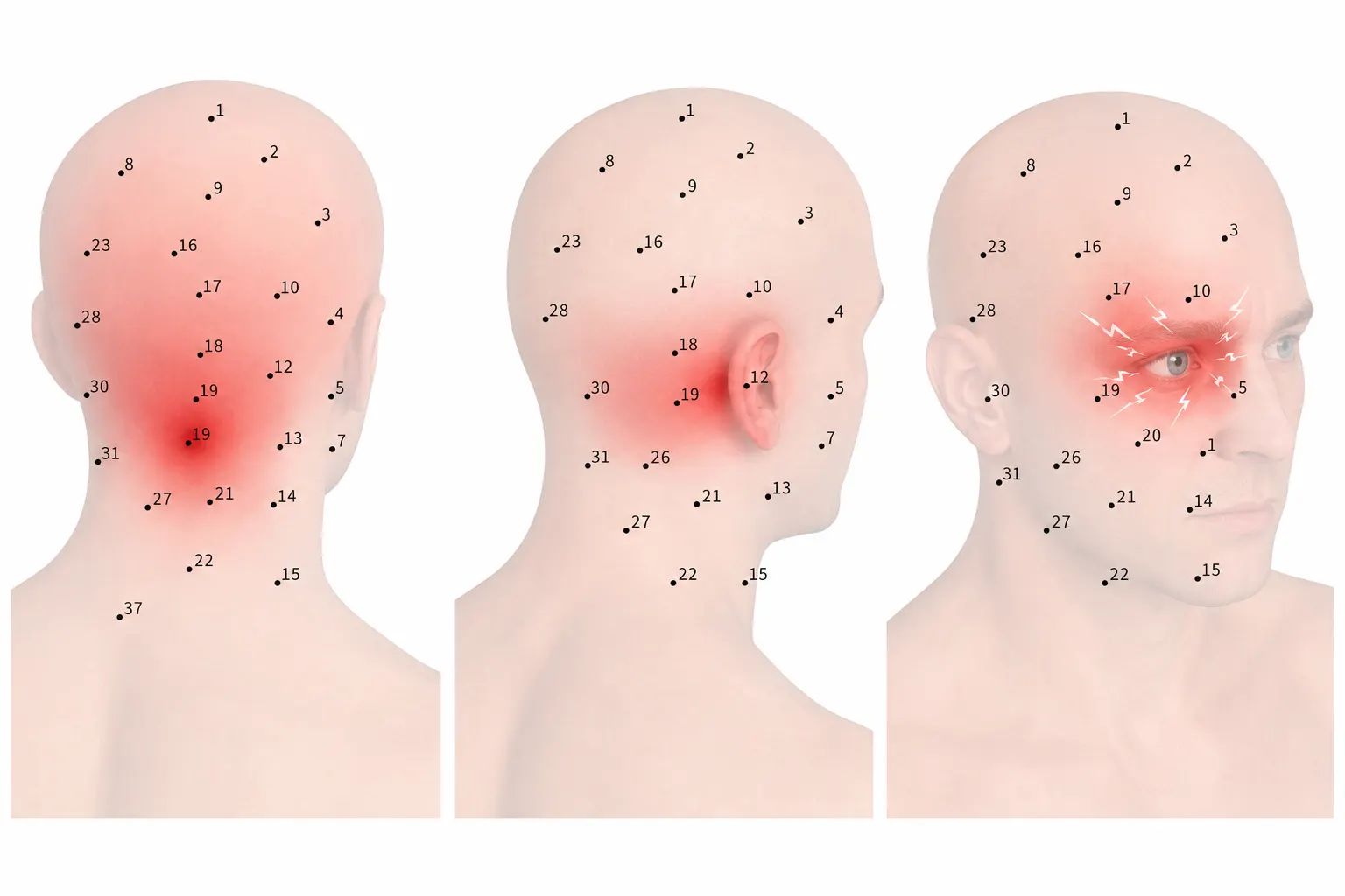
Sharp, shooting, often described as "electric" pain. Between attacks, a dull sensation of pressure often remains in the back of the head.
Beginning at the base of the skull, radiating across the crown, temple — in more severe cases up to behind the ipsilateral eye (so-called convergent nociceptive pathways).
Typical is tenderness to pressure over the occipital nerve at the base of the skull. Triggers are often neck rotation, prolonged forward leaning, or pressure on the back of the head (pillow, helmet).
Whiplash injuries, prolonged screen time, constriction situations in the area of the upper cervical spine and neck muscles — the result is mechanical irritation of the nerve fibres.
According to ICHD-3 (13.4), the diagnosis requires paroxysmal, stabbing pain in the distribution of the greater/lesser/third occipital nerve, with tenderness to pressure and (temporary) response to a diagnostic nerve block. The most frequent cause is compression of the greater (approx. 90% of cases) or lesser (approx. 10%) occipital nerve; individual diagnostic blocks have false-positive rates of up to 40%, which is why a second block is useful for confirmation.
ICHD-3, Section 13.4 Occipital neuralgia, ichd-3.org. — Choi I, Jahnke S. Occipital Neuralgia — StatPearls, NBK538281. — Vanterpool CS et al. Ultrasound-Guided Greater Occipital Nerve Blocks and PRFA for Occipital Neuralgia. PMC3833045.The greater auricular nerve is a sensory branch of the upper cervical plexus (C2/C3). It supplies the skin over the auricle, mastoid, angle of the mandible, and parotid gland. Its neuralgia is often confused with ear, temporomandibular joint, or trigeminal complaints — patients travel specifically to Düsseldorf for diagnosis and therapy.
Persistent burning or pulling, interspersed with shooting, seconds-long stabs. Often accompanied by sensory disturbances, tingling, or numbness in the supply area.
In front of and behind the auricle, over the mastoid process, at the angle of the jaw, and laterally on the cheek/neck — exactly in the nerve's supply area.
Tinel-like sign over the course of the nerve on the sternocleidomastoid. Provocation through head rotation, side-lying, tight collars, or pressure in the surgical area.
Common triggers include neck surgeries (parotid, carotid, lymph nodes), blunt neck trauma, as well as prolonged pressure — e.g., sleeping on the side, heavy helmets, tight clothing.
Neuralgia of the greater auricular nerve (C2/C3) is rare and documented in the literature predominantly as case series. In the largest published series (13 cases), sufferers mostly responded clearly to diagnostic nerve blocks; for therapy-refractory courses, peripheral nerve stimulators were also used. Standard neuropathic medication showed no sufficient effect in several cases.
Duvall JR, Garza I, Kissoon NR et al. Great Auricular Neuralgia: Case Series. Headache 2020;60(1):247–258. PMID 31749202. — Robertson CE, Garza I. Great auricular neuralgia: case series. J Headache Pain 2013;14(Suppl 1):P161. — Christ S et al. Identification of the great auricular nerve by ultrasound imaging and transcutaneous nerve stimulation. Anesth Analg 2012;114:1128–1130.Occipital, auricular, and trigeminal nerves are anatomically close to each other and are often confused in practice. This overview shows the clinically most important differences.
Classification is done clinically — history, targeted pressure and provocation findings, and if necessary, a diagnostic nerve block secure the diagnosis and define the therapeutic goals.
Axomera (percutaneous bioelectric current stimulation, PBCS) generates controlled static electric fields in the treatment area within the range of naturally occurring potentials — addressing the "electrical dimension" of inflammation and pain.
For whiplash injury and occipital neuralgia, we treat near the nerve at the upper cervical spine, for greater auricular neuralgia along the course of the neck, for SUNCT in the eye-forehead area of the trigeminal nerve. For patients, the treatment feels similar to acupuncture; technically, it is different — fine probes, low physiological currents, microprocessor-controlled.
Inflammation has an electrical dimension. Axomera starts exactly there.
Axomera is an internationally patented bioelectrical procedure.
How Axomera therapy works →Conventionally, pain medication, manual therapy, anticonvulsants (for neuralgias), nerve blocks, and injections are used. We know these paths and their limits from our orthopaedic pain medicine practice.
The goal is to break a chronic course — for headache after whiplash injury just as for occipital and greater auricular neuralgia. Statements on the number of sessions remain guide values from our experience.
We state this consciously as a goal. How much relief can be achieved in individual cases depends on the individual course.
Developer of Axomera therapy · Medical Director
As the developer of Axomera therapy, he oversees the indication and is involved in your treatment personally upon request. His experience in conservative pain medicine informs the planning of your treatment series.
Doctor, orthopaedics & trauma surgery (in specialist training)
Performs your Axomera treatment and accompanies you throughout the entire treatment series. He reviews your findings and assesses whether the procedure is suitable for you. He also participates in the technical development.
Doctor, Specialist in Orthopaedics and Trauma Surgery
Organises your previous findings, performs Axomera diagnostics, and accompanies you throughout the entire treatment series. He reviews your findings and, based on his experience, assesses whether the procedure is suitable for you.
Three ways to reach us, around the clock, directly here on the site. For each, you can upload doctor's letters, findings, and MRI images at the same time — encrypted into your file, reviewed by a doctor before we speak.
Ten minutes with a doctor. Free of charge.
Medical assessment via video.
Examination, Axomera diagnostics, plan, costs.
Prefer personal contact? Call: +49 211 866880
Our practice treats exclusively by prior appointment.
The telephone initial assessment is not a treatment and does not replace an examination. Whether Axomera is suitable for you will be decided after a medical examination.