axomera
Shoulder pain · Düsseldorf

Small inflammation. Big effect. Precisely localised.

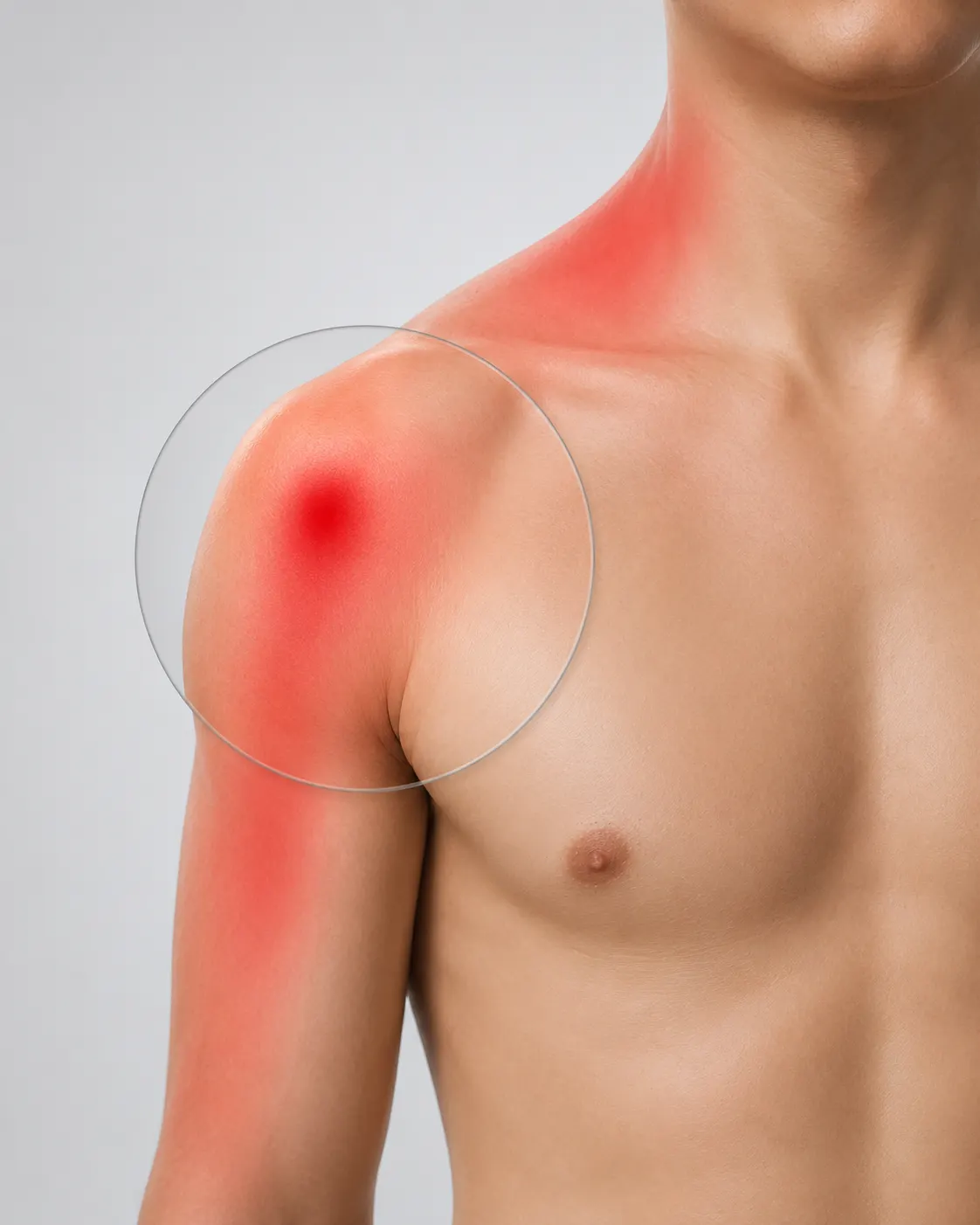
Shoulder pain is persistent and has many possible causes. As a reference centre for Axomera therapy, we look for the painful region — whether rotator cuff, impingement or calcific shoulder — often the smallest inflammations with maximal impairment — and treat them bioelectrically, if necessary in combination with acupuncture and other procedures.

Medical content overseen by Prof. Dr. med. Albrecht Molsberger · reviewed 10/2026

Magnifying glass over the painful shoulder region
Clinical picture

Many causes — usually protracted.

Shoulder pain have many causes — impingement syndrome, irritation or partial damage to the rotator cuff, calcific shoulder or bursitis.

It is characteristic that shoulder diseases are often protracted and drag on for many months; the longest possible conservative treatment is usually sensible.

Your medical team

Who treats you.

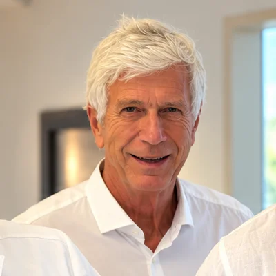
Medical team at Axomera Practice Düsseldorf: Dr. med. Philipp Schröder, Prof. Dr. med. Albrecht Molsberger and Jonas Becker
From left to right
  • Dr. med. Philipp SchröderDoctor, orthopaedics & trauma surgery (in specialist training)
  • Prof. Dr. med. Albrecht MolsbergerSpecialist in orthopaedics
    Medical Director
  • Jonas BeckerSpecialist in orthopaedics and trauma surgery

At the reference centre for Axomera therapy, you are treated by a medical team led by Prof. Molsberger.

Patients

What patients report.

Typical findings

From our patient consultations and the medical reports submitted.

  • Complaints usually since several months to over a year
  • Pain when lifting and turning the arm
  • Nocturnal pain when lying on the shoulder
  • Restricted movement
  • Often multiple doctors and pre-treatments. Surgery recommended.
Previous therapy

Almost always in this order.

  1. Rest and painkillers (NSAIDs)
  2. Physiotherapy (strengthening the rotator cuff)
  3. Cortisone injection (subacromial)
  4. Shockwave (mainly for calcific shoulder)
  5. Surgery (e.g. arthroscopy / decompression / tendon repair)
MRI findings of the shoulder joint

Not every MRI finding explains the pain.

Rotator cuff changes and supraspinatus partial ruptures in MRI are in many cases age-related incidental findings without clinical correlation. The prevalence of asymptomatic rotator cuff changes increases continuously with age:

7 %under 20 years
26 %41–60 years
50 %61–80 years
56 %from 80 years

In population-based ultrasound studies, an additional 11–17% of completely asymptomatic shoulders have demonstrable full-thickness ruptures of the rotator cuff. A tendon tear visible on MRI is therefore not automatically the cause of the complaints — clinical examination, complaint pattern and progression are often more informative than the image. Before any decision for surgery, the agreement between image findings and clinical complaint pattern should therefore be carefully examined.

Teunis T et al. Prevalence of rotator cuff disease with increasing age, n=6,112 shoulders. J Shoulder Elbow Surg 2014;23(12):1913–1921. — Rotator Cuff Imaging Abnormalities in Asymptomatic Shoulders: SR, 53 studies. JOSPT 2025. — Yamamoto A et al. Prevalence of symptomatic and asymptomatic rotator cuff tears in the general population. PMC3768248.

Biceps tendon

Biceps tendon pain — often misinterpreted.

A “sensitive point” over the long biceps tendon in the area of the anterior shoulder is among the most frequent palpation findings in shoulder pain — but it is diagnostically far less informative than is often assumed. Isolated pathologies of the biceps tendon are relatively rare in imaging studies at about 25%:

42 %Biceps tendon and rotator cuff intact
27 %isolated biceps tendon pathology
14 %isolated rotator cuff pathology
18 %combined pathology of both structures

The classic anterior shoulder pain radiating into the upper arm is also not specific — it just as well arises in rotator cuff pathology, SLAP lesions, subacromial impingement or a cervical nerve root irritation (C5/C6). Furthermore, histological examinations show that the classic “biceps tendinitis” is usually not an acute inflammation, but a degenerative tendinopathy — a finding that relativises the expectations of anti-inflammatory drugs. A positive palpation finding or an MRI signal at the biceps tendon is therefore never an isolated reason for an operation — in the majority of patients, the cause of the complaints lies in the surrounding shoulder complex.

Cheng L et al. Humeral head migration and biceps tendon pathology, 3T MRI cohort, n=79. PMC9446281. — Redondo-Alonso L et al. Supraspinatus and long biceps tendon, SR. BMC Musculoskelet Disord 2014;15:377. PMC4247626. — Krupp RJ et al. Proximal biceps tendinitis and tendinopathy. StatPearls 2023. NBK533002. — Ellman MB, Yanke AB, Cole BJ. Diagnosis and management of biceps tendinitis. J Am Acad Orthop Surg. — Kircher J et al. Surgical treatment trends of LHB pathology 2010–2019. PMC12047552.

Standard therapies in focus

What standard therapies really achieve.

Conservative therapy: medication and physiotherapy.

NSAIDs and painkillers alleviate acute shoulder pain in the short term, but do not change the long-term course; for cortisone injections, a short-term effect in the first 6–8 weeks is proven, thereafter no advantage over placebo, and repeated injections can impair tendon quality. According to current evidence, frozen shoulder is not understood as a primarily mechanical, but as a neuroimmunological-fibrotic event with fibroblast activation and cytokine dysregulation (IL-1β, IL-6, TNF-α) — the capsule becomes inflamed and fibroses without a mechanical cause. In this constellation, a consistently conducted conservative therapy over months is the rule.

Lo CS et al. Combination vs. single physio for RCRSP, network MA. J Clin Med 2025. PMC12250685. — Ryan V et al. Pathophysiology frozen shoulder, SR. BMC Musculoskelet Disord 2016;17:340. — Akbar M et al. Fibroblast & Inflammation. PLoS ONE 2019;14:e0215301.

Acupuncture.

In chronic shoulder pain, the GRASP study (n=424, patient-blinded, BMBF-funded) shows in direct comparison that 15 acupuncture treatments over 6 weeks are statistically significantly superior to a conventional standard therapy (NSAIDs, physiotherapy, cortisone): responder rate 65% with acupuncture versus 37% with conventional therapy (p<0.01, 3 months after end of therapy). Current meta-analyses confirm this effect in rotator cuff-related shoulder pain and in the postoperative rehabilitation process; acupuncture is, based on evidence, among the most effective conservative options for chronic shoulder complaints.

Molsberger AF et al. GRASP: Responder Verum 65% vs. conventional 37% vs. Sham 24% (p<0.01), n=424. Pain 2010;151:146–154. PMID 20655660. — Chen JH et al. Acupuncture in RC rehab, MA of 18 RCTs (n=376). Front Surg 2025. PMC12454321.

Shockwave therapy.

Extracorporeal shockwave therapy (ESWT) has a clearly defined indication for shoulder conditions: calcific tendonitis (Tendinosis calcarea) with symptomatic calcium deposits. In this condition, ESWT can fragment the calcium deposit and accelerate resorption; the evidence for pain reduction and functional improvement is proven in several randomised studies. For other shoulder pathologies — rotator cuff tendinopathy without calcification, subacromial pain syndrome, frozen shoulder — the study situation is heterogeneous and a specific benefit is less convincing than for calcific tendonitis. Treatment is outpatient and minimally invasive, but in cases of incorrect indication, it can also worsen the pain progression. Shockwave therapy has its place for symptomatic calcium deposits — it should not be used routinely for other shoulder problems.

Bannuru RR et al. High-energy ESWT for chronic calcific tendinitis of the shoulder: Meta-analysis. Ann Intern Med 2014. — Xue X et al. ESWT for rotator cuff injury: SR protocol. PLoS ONE 2024. PMC11078342.

Operation: subacromial decompression — debunked by placebo-controlled trials.

The most common shoulder arthroscopy is subacromial decompression — a procedure whose efficacy has been fundamentally questioned by two landmark placebo-controlled studies: the CSAW study (Lancet 2018, n=313) and the FIMPACT study (BMJ 2018, 5-year follow-up 2020, n=210) showed no significant difference in outcome in randomised comparisons between subacromial decompression, placebo arthroscopy, and physiotherapy — neither after 12 months nor after 5 years. MRI studies also prove that rotator cuff tears are often age-related, asymptomatic incidental findings: the prevalence increases from 6.7% in those under 20 to over 50% in those over 80 (Teunis meta-analysis, n=6,112 shoulders), and in population studies, 11–17% of asymptomatic shoulders have sonographically detectable full-thickness ruptures. A supraspinatus tear visible on an MRI is therefore not automatically the cause of pain — clinical examination and clinical progression are more decisive than the imaging findings. Shoulder arthroscopies are indicated surgically in cases of traumatic ruptures with a clinical correlate, functionally relevant deficits, and after several months of unsuccessful conservative therapy. Before any elective shoulder surgery, the discrepancy between imaging findings and complaints, as well as the effectiveness of conservative procedures — particularly the acupuncture proven in the GRASP study — should be evaluated.

Beard DJ et al. CSAW: placebo-controlled RCT. Lancet 2018;391:329–338. — Paavola M et al. FIMPACT: placebo-surgery RCT. BMJ 2018;362:k2860. — Paavola M et al. FIMPACT 5-year follow-up. BJSM 2020. PMC7788208. — Teunis T et al. SR of rotator cuff disease prevalence, n=6,112. J Shoulder Elbow Surg 2014. — Yamamoto A et al. Symptomatic and asymptomatic rotator cuff tears. PMC3768248.

Where we make the difference

Cortisone injections under the acromion often only work for a few weeks, and for many findings, arthroscopy is not necessarily the next logical step.

Axomera for shoulder pain.

Under the acromion, the tendons of the rotator cuff and a bursa rub against each other with every arm movement; if the strain persists, tendon remodeling falls behind and the inflammation becomes self-perpetuating. These structures are located directly under the acromion and are easily accessible from the outside.

Axomera targets this region: computer-controlled electrical signals in the dimension of cellular electrical fields are placed with millimeter precision at tendon attachments and the bursa under the acromion. The goal is to shift the disturbed tendon remodeling back in favour of healing. This neurophysiological hypothesis is also supported empirically by many patient progressions (real-world data), not by controlled studies; as with other indications, around 20–30% of those treated do not respond.

How inflammation and pain are connected →
„
Athletes do not just want to be pain-free. They want to be able to load the joint again — that is what we measure ourselves by.
Dr. med. Philipp Schröder
— Dr. Philipp Schröder · Ultrasound-guided probe placement, co-developer of the Axomera therapy

Axomera is an internationally patented bioelectrical procedure.

How Axomera therapy works →
Our approach

How we proceed with you.

Every therapy needs its own examination — and the Axomera diagnostics is the prerequisite for targeted Axomera therapy.

Axomera diagnostics
  1. Secure the diagnosis and check the influence of taken medication
  2. Millimeter-precise examination — locate pain points in the irritated tendon and soft tissue region of the shoulder
Axomera therapy
  1. 4–6 Axomera treatments, precisely at these points, often in combination with acupuncture.
  2. Goal: significant reduction of pain and improvement of mobility
  3. In favorable courses: shortening of the disease progression with full functional capacity of the shoulder in everyday life. Avoidance of surgery.

Aims & course of treatment. We cannot guarantee a specific result — the progression is individual. Depending on the course of the condition, fewer or more treatments may be required.

Axomera treats the irritated tendon and soft tissue region of the shoulder bioelectrically. It works at a level deeper than painkillers, injections, and shockwave. Axomera directly modulates the body's own control of inflammation and healing — and is combined with acupuncture and other procedures if required.

Patients report

Individual cases.

After just three weeks of shoulder pain, an arthroscopic operation was recommended to me — the supraspinatus tendon allegedly had no more space and was torn. I decided against it. Instead: Axomera, acupuncture, physiotherapy, and four months of patience. Today my shoulder feels completely healthy again — without surgery.
Individual case

Four months of patience instead of arthroscopy.

E. H., patient — documented individual case from the practice.

Documented individual cases, with patient consent. Individual outcomes differ.

Is Axomera right for me? Initial telephone assessment →
Your treatment team

Who treats you.

Your practitioners
Prof. Dr. med. Albrecht Molsberger

Prof. Dr. med. Albrecht Molsberger

Developer of Axomera therapy · Medical Director

As the developer of Axomera therapy, he oversees the indication and is involved in your treatment personally upon request. His experience in conservative pain medicine informs the planning of your treatment series.

Career history
  • Specialist in Orthopaedics; additional qualifications in Sports Medicine, Acupuncture, Naturopathy
  • Habilitation 2005, adjunct professorship for Orthopaedics at Ruhr University Bochum
  • Conception and management committee of the GERAC studies (German Acupuncture Trials)
  • Development of Axomera therapy since 2008
Dr. med. Philipp Schröder

Dr. med. Philipp Schröder

Doctor, orthopaedics & trauma surgery (in specialist training)

Performs your Axomera treatment and accompanies you throughout the entire treatment series. He reviews your findings and assesses whether the procedure is suitable for you. He also participates in the technical development.

Career history
  • Doctor, orthopaedics & trauma surgery (in specialist training); medical degree, Heinrich Heine University Düsseldorf, 2020
  • Doctorate magna cum laude, Ruhr University Bochum (2021): Pain localisation in cluster headache
  • Expert courses in Ultrasound in Pain Medicine (DEGUM curriculum, level I); Expert course in Sports Medicine
  • Technical co-development of Axomera therapy; co-founder of Columbus Health-Products GmbH
Further practitioner
Jonas Becker

Jonas Becker

Doctor, Specialist in Orthopaedics and Trauma Surgery

Organises your previous findings, performs Axomera diagnostics, and accompanies you throughout the entire treatment series. He reviews your findings and, based on his experience, assesses whether the procedure is suitable for you.

Contact · Online reception

Clarify first. Then travel.

Three ways to reach us, around the clock, directly here on the site. For each, you can upload doctor's letters, findings, and MRI images at the same time — encrypted into your file, reviewed by a doctor before we speak.

01 · Telephone

Briefly check if it's a fit.

Ten minutes with a doctor. Free of charge.

  • ·For those who are unsure
  • ·Upload findings beforehand
  • ·No obligation
Select phone appointment →
02 · Video

Consultation on findings without the travel.

Medical assessment via video.

  • ·For patients traveling from further away
  • ·Second opinion before making a decision
  • ·According to GOÄ, amount visible upon booking
Book video appointment →
03 · Practice

Everything in one appointment.

Examination, Axomera diagnostics, plan, costs.

  • ·For all who wish to visit
  • ·The direct route for distances up to about 100 km
  • ·Bring or upload findings
Book appointment →

Prefer personal contact? Call: +49 211 866880

Our practice treats exclusively by prior appointment.

The telephone initial assessment is not a treatment and does not replace an examination. Whether Axomera is suitable for you will be decided after a medical examination.