axomera
Knee pain · Düsseldorf

First, understand where the pain comes from. Then, treat it.

The knee rarely hurts where the X-ray and MRI show damage. As an Axomera therapy reference centre in Düsseldorf, we look for the structure that is causing the pain — and we treat it.

Medical content overseen by Prof. Dr. med. Albrecht Molsberger · reviewed 10/2026

Structurally, everything is supposed to be fine — but I cannot walk.

Frequent patient statement · Sport overuse

A characteristic, recurring patient statement from our consultation hours.

Three typical paths

How patients come to us.

The athlete

The knee that no longer keeps up.

Runners, footballers, tennis players. Pain at the front of the kneecap, inside the joint space, or at the tendon attachment — creeping in with training load. Imaging is often unremarkable.

"Structurally, everything is supposed to be fine, but I cannot walk."

The 50+ patient

The worn-out knee.

Pain during exertion — walking downstairs, standing up, after sitting for a long time. The X-ray shows osteoarthritis, a narrower joint space. The recommendation: wait and see, painkillers, eventually an artificial joint.

"I was told there was nothing more to be done except surgery."

After the artificial joint

Pain despite a correct prosthesis.

The endoprosthesis is positioned correctly, the check-up is unremarkable — yet the knee still hurts. At the front, on the inside, sometimes burning or with a hypersensitive point.

"The surgeon says everything is technically perfect. But it still hurts."

Clinical picture

A symptom, not a diagnosis.

Knee pain can originate from inside the joint, from the surrounding soft tissue, from the bone beneath the cartilage, or from an irritated nerve — and sometimes it even projects into the knee from the hip or lower back.

Therefore, we do not start by asking "How severe is the damage?", but: Which structure is creating the pain?

Your medical team

Who treats you.

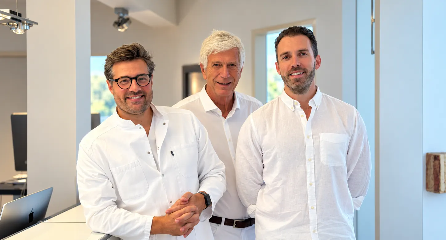
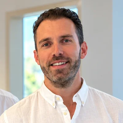
Medical team at Axomera Practice Düsseldorf: Dr. med. Philipp Schröder, Prof. Dr. med. Albrecht Molsberger and Jonas Becker
From left to right
  • Dr. med. Philipp SchröderDoctor, orthopaedics & trauma surgery (in specialist training)
  • Prof. Dr. med. Albrecht MolsbergerSpecialist in orthopaedics
    Medical Director
  • Jonas BeckerSpecialist in orthopaedics and trauma surgery

At the reference centre for Axomera therapy, you are treated by a medical team led by Prof. Molsberger.

Patients

What patients report.

Typical findings

From our patient consultations and the medical reports submitted.

  • Pain dependent on exertion — walking downstairs, standing up, after sitting for a long time, or with training load
  • Pain at the front of/behind the kneecap, inside the joint space, or at the tendon attachment
  • Starting pain after rest that "walks off" but returns with overuse
  • Persistent pain despite unremarkable imaging — or despite a technically correct endoprosthesis
  • A circumscribed, touch-sensitive point — an indication of a nerve component
Previous therapy

The established stages — conservative and surgical.

  1. Waiting & rest. Sensible for many acute irritation states — no solution for persistent complaints.
  2. Painkillers and anti-inflammatories. They dampen the signal, not the cause — not intended for long-term use.
  3. Physiotherapy & muscle building. Only works reliably once the source of pain in the joint has been calmed.
  4. Injections. Cortisone, hyaluronic acid, biological procedures (PRP, autologous serum) — the data basis varies depending on the procedure.
  5. Arthroscopy / Surgery. Indicated for genuine mechanical causes; limited benefit for pure wear-related meniscus issues.
  6. Artificial joint (endoprosthesis).
Where we make the difference

Hyaluronic acid and cortisone injections often only work for a few months, and an artificial joint is only an option for many patients after years.

Axomera for knee pain and knee osteoarthritis.

In the osteoarthritic knee, it is primarily the nerve-rich soft tissues — the joint capsule, synovial membrane, and the bone directly beneath the cartilage — that generate the pain, not the loss of cartilage itself. These structures are located near the surface around the joint and are precisely reachable from the outside.

Axomera targets these soft tissue structures: computer-controlled electrical signals at the dimension of cellular electrical fields are placed millimetre-precisely at the capsule, synovial membrane, and subchondral bone. The goal is to shift the inflammation-regeneration balance in favour of healing — this does not reverse the cartilage wear itself.

How inflammation and pain are connected →
„
Inflammation has an electrical dimension. Axomera starts exactly there.
Prof. Dr. Albrecht Molsberger
— Prof. Dr. Albrecht Molsberger · Developer of Axomera therapy, medical director

Axomera is an internationally patented bioelectrical procedure.

How Axomera therapy works →
Our approach

How we proceed with you.

Every therapy needs its own examination — for us, this includes the Axomera diagnostics and the Axomera therapy.

Axomera diagnostics
  1. Locate the pain-generating structure — peripatellar soft tissues, tendon attachment, subchondral bone, or an irritated nerve. Imaging shows damage; it does not reliably show the location of the pain.
  2. Separate pain and irritation components — how much originates from mechanical load, inflammation, and a nerve component.

This examination complements imaging and medical diagnosis — it does not replace them.

Axomera therapy
  1. Fine electrical probes are placed precisely at the structure identified as pain-generating.
  2. A short series of sessions over a few weeks — number and frequency individual. Goal: reduce inflammation and pain.
  3. Connect with active rehabilitation — once the cause has been calmed, muscle building works reliably.

Aimed goals. We cannot guarantee a specific result — progressions are individual.

According to cell biological basic research, weak, endogenous electrical fields control cell migration, inflammation, and healing — Axomera therapy addresses this level.

A deeper look

Five questions that are almost always answered incorrectly regarding the knee.

Knee pain does not come from worn-out cartilage.

It sounds obvious: The X-ray shows worn cartilage, so the cartilage must hurt. But that is simply not true — cartilage has no nerves; it cannot generate pain. A medic demonstrated this impressively by having his own knee palpated from the inside without anaesthetic: The cartilage was silent, while the joint lining, Hoffa's fat pad, and capsule reacted with severe pain. Where does the pain come from, then? From the well-innervated structures around the cartilage — joint lining, fat pad, lateral support apparatus, bone directly beneath the cartilage. Severe cartilage damage in the image and barely any complaints, or strong pain with nearly intact cartilage: Damage in the image and the site of pain are two different things. This is exactly why we search for the pain-generating structure first.

Dye SF et al. Conscious neurosensory mapping of the internal structures of the human knee without intraarticular anesthesia. Am J Sports Med 1998. — Sanchis-Alfonso V, Roselló-Sastre E. Immunohistochemical analysis of the lateral retinaculum. Am J Sports Med 2000. — Adatia A et al. Osteoarthritis of the knee and hip, Part I. J Pharm Pharmacol 2012.

"Chondropathia patellae" — a name that misses the cause.

The anterior knee pain already carries the word cartilage ("Chondro") in its name. Yet the pain is not in the cartilage of the kneecap, but in the lateral support apparatus, in the fat pad with the joint lining, and in the bone beneath the joint surface. The term describes an imaging finding and thus names the wrong structure. For treatment, this difference is decisive: Anyone wanting to "repair" the cartilage is targeting a tissue that does not hurt at all. Anyone who calms the actual pain-generating soft tissues is addressing the cause. That is why we differentiate so precisely at the front of the knee between cartilage findings and the location of the pain.

Bentley G, Dowd G. Current concepts of etiology and treatment of chondromalacia patellae. Clin Orthop Relat Res 1984. PMID 6383677. — Sanchis-Alfonso V, Roselló-Sastre E. Am J Sports Med 2000.

Meniscus: when an operation helps — and when it does not.

A meniscus tear is one of the most common incidental findings in a knee MRI and is often operated on, even though the best available evidence warns against it. In a sham-controlled study, arthroscopic partial removal in degenerative (wear-related) meniscus tears showed no benefit compared to the sham procedure — the tear is usually a side effect of ageing here, not the source of pain. It is different with a genuine mechanical problem: a trapped, displaced fragment, a blocked knee, or a fresh injury tear in a younger, active person — here surgery is correct, and if the meniscus is operated on, the rule today is: preserve as much as possible (stitch) instead of removing. In short: Wear-tear without blockage → conservative. Injury tear or genuine blockage → operate, meniscus-preserving.

Sihvonen R et al. Arthroscopic partial meniscectomy versus sham surgery for a degenerative meniscal tear. N Engl J Med 2013;369:2515–24. — Lee SH et al. Revisiting APM for degenerative tears, Meta-Analyse. Clin J Sport Med 2020. — Stein T et al. Long-term outcome after arthroscopic meniscal repair versus partial meniscectomy. Am J Sports Med 2010.

Why the thigh muscle becomes weaker — and what needs to be treated first.

When the knee hurts, the thigh muscles waste away; the usual advice is then: build up the muscle. That is not wrong — but the order is often not right. The muscle weakness is usually a consequence of the knee pain, not its cause. Technically, the phenomenon is called arthrogenic muscle inhibition: The irritated joint sends altered signals that reflexively throttle muscle control. It is difficult to strengthen a muscle that your own nervous system is currently downregulating. This results in a clear sequence: first calm the source of pain and irritation in the joint, then build up in a targeted manner. That is why we do not use physiotherapy against, but after or alongside the treatment of the pain cause — not an either-or.

Forelli F et al. Rethinking the Assessment of Arthrogenic Muscle Inhibition After ACLR. J Clin Med 2025. — Smith MJ et al. Quadriceps Muscle Deficits following ACL Injury. Sports Med 2025. — Alammari A et al. Hip abductor/rotator strengthening in PFP, SR & Meta-Analyse. J Back Musculoskelet Rehabil 2023.

Not all knee pain is wear or overuse.

Osteoarthritis and mechanical incorrect or overuse explain most unilateral knee pains — but not all. Sometimes a hip or the lower back projects its pain into the knee. Sometimes an irritated nerve is behind it, for example after surgery, if a fine skin nerve on the inside of the knee is trapped or has formed a neuroma. And there are bone reactions beneath the joint surface that look like osteoarthritis in the image, but are independent causes. Rare, but important to rule out: infection and tumour. Before we accept "osteoarthritis" or "overuse" as an explanation, we rule these out.

Malghem J et al. Subchondral insufficiency fracture / SONK. Insights Imaging 2023. — Yang SR et al. Neuralgia des R. infrapatellaris n. saphenus als Ursache anteromedialen Knieschmerzes nach Knie-OP. Eur Radiol 2021.

Patients report

Documented progress.

After an arthroscopically operated meniscus tear, residual complaints remained at a small point on the knee joint capsule. Two Axomera therapies were carried out there — I have been symptom-free ever since.
After arthroscopy · 48

Symptom-free ever since.

Patient, 48 — marked individual progress.

Documented individual cases, with patient consent. Individual outcomes differ.

Is Axomera right for me? Initial telephone assessment →
Your treatment team

Who treats you.

Your practitioners
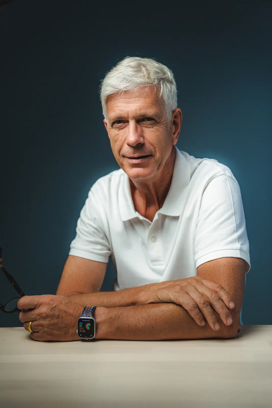
Prof. Dr. med. Albrecht Molsberger

Prof. Dr. med. Albrecht Molsberger

Developer of Axomera therapy · Medical Director

As the developer of Axomera therapy, he oversees the indication and is involved in your treatment personally upon request. His experience in conservative pain medicine informs the planning of your treatment series.

Career history
  • Specialist in Orthopaedics; additional qualifications in Sports Medicine, Acupuncture, Naturopathy
  • Habilitation 2005, adjunct professorship for Orthopaedics at Ruhr University Bochum
  • Conception and management committee of the GERAC studies (German Acupuncture Trials)
  • Development of Axomera therapy since 2008
Dr. med. Philipp Schröder

Dr. med. Philipp Schröder

Doctor, orthopaedics & trauma surgery (in specialist training)

Performs your Axomera treatment and accompanies you throughout the entire treatment series. He reviews your findings and assesses whether the procedure is suitable for you. He also participates in the technical development.

Career history
  • Doctor, orthopaedics & trauma surgery (in specialist training); medical degree, Heinrich Heine University Düsseldorf, 2020
  • Doctorate magna cum laude, Ruhr University Bochum (2021): Pain localisation in cluster headache
  • Expert courses in Ultrasound in Pain Medicine (DEGUM curriculum, level I); Expert course in Sports Medicine
  • Technical co-development of Axomera therapy; co-founder of Columbus Health-Products GmbH
Further practitioner
Jonas Becker

Jonas Becker

Doctor, Specialist in Orthopaedics and Trauma Surgery

Organises your previous findings, performs Axomera diagnostics, and accompanies you throughout the entire treatment series. He reviews your findings and, based on his experience, assesses whether the procedure is suitable for you.

Safety warning. A suddenly blocked knee that can no longer be straightened, an acutely severely swollen, overheated, and reddened joint, or rapidly progressing rest pain should be clarified immediately by a specialist.

Contact · Online reception

Clarify first. Then travel.

Three ways to reach us, around the clock, directly here on the site. For each, you can upload doctor's letters, findings, and MRI images at the same time — encrypted into your file, reviewed by a doctor before we speak.

01 · Telephone

Briefly check if it's a fit.

Ten minutes with a doctor. Free of charge.

  • ·For those who are unsure
  • ·Upload findings beforehand
  • ·No obligation
Select phone appointment →
02 · Video

Consultation on findings without the travel.

Medical assessment via video.

  • ·For patients traveling from further away
  • ·Second opinion before making a decision
  • ·According to GOÄ, amount visible upon booking
Book video appointment →
03 · Practice

Everything in one appointment.

Examination, Axomera diagnostics, plan, costs.

  • ·For all who wish to visit
  • ·The direct route for distances up to about 100 km
  • ·Bring or upload findings
Book appointment →

Prefer personal contact? Call: +49 211 866880

Our practice treats exclusively by prior appointment.

The telephone initial assessment is not a treatment and does not replace an examination. Whether Axomera is suitable for you will be decided after a medical examination.